Submitted by admin on Sat, 09/20/2008 - 21:10
tissue_organ_import:
brain, cerebellum
Morphologic diagnosis:
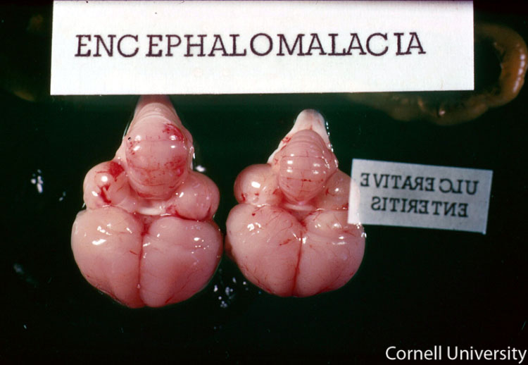
Cerebellum: Severe edema and mild hemorrhage
Clinical description:
On post-mortem examination, gross lesions may be observed on the brains of birds with encephalomalacia. The most common lesions are usually found on the cerebellum. The cerebellum of the affected bird on the left shows swelling, blunting of the ridges, and small petechial hemorrhages on its surface. The normal brain on the right is shown for comparison.
Pathologic description:
This picture shows the brains of two birds. The brain on the right is normal. The cerebellum of the bird on the left is markedly swollen, the normal ridges are obscured, and the surface of the tissue is covered by multiple variably-sized petechiae.
Record number:
7102
Case number:
Unknown
Breed:
White Leghorn
Clinical form:
Unknown
Infection type:
Unknown
Housing/mgmnt type:
Select One
Priority:
1
Rights:
© Cornell University
Image source URL:
http://cidc.library.cornell.edu/vet_avian/images/VIT E-SELENIUM adjusted/VIT E-SELENIUM - 056A.jpg
Etiology:
Exam findings:
Tissues and organs:
Asset type:
Species:
Image:

