Submitted by admin on Sat, 09/20/2008 - 21:10
tissue_organ_import:
trachea
Morphologic diagnosis:
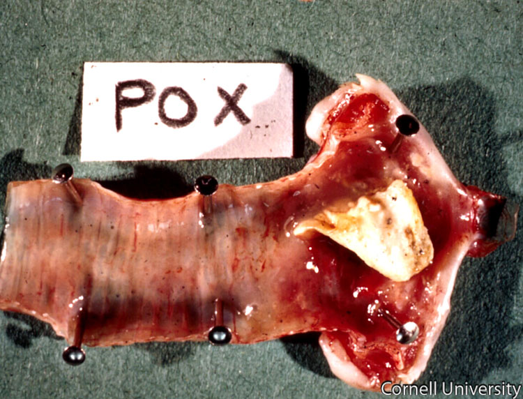
Trachea: Locally extensive fibrinonecrotic tracheitis with multifocal petechia.
Clinical description:
Diphtheritic (wet) form of avian pox. On post-mortem examination, if these diptheritic membranes are removed, bleeding erosions are sometimes found beneath these membranes.
Pathologic description:
The trachea has been opened to reveal the mucosal surface. Within the cranial portion of the esophagus (right side of picture) there is a large aggregate of dry, pale tan, friable material. The material overlies a bright red and wet section of mucosa. Small pinpoint red foci are scattered along more distal portions of the tracheal mucosa.
Record number:
7671
Case number:
Unknown
Clinical form:
Unknown
Infection type:
Unknown
Housing/mgmnt type:
Select One
Priority:
1
Image source URL:
http://cidc.library.cornell.edu/vet_avian/images/POX Adjusted/POX-053A.jpg
Etiology:
Exam findings:
Asset type:
Species:
Image:

