Submitted by admin on Sat, 09/20/2008 - 21:10
tissue_organ_import:
embryo
Morphologic diagnosis:
Chorioallentoic membrane: Multifocal hyperplasia.
Clinical description:
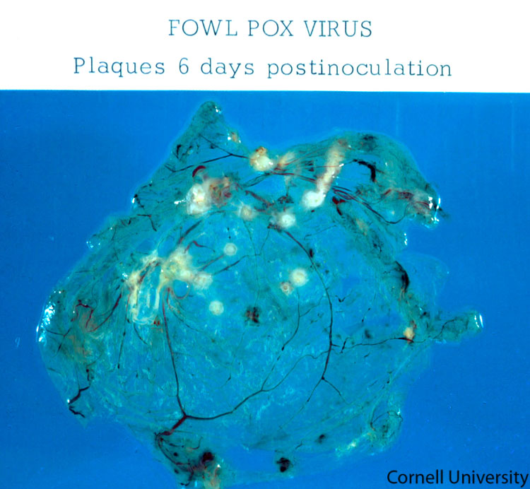
In laboratory diagnosis of avian pox, chicken embryo inoculation is one of the best methods to identify typical lesions induced by this virus. Here, white opaque pock lesions have developed 6 days post-inoculation on the chorioallantoic membrane (CAM) of a chicken embryo inoculated with poxvirus.
Pathologic description:
The normally transparent tissues of the chorioallantoic membrane are disrupted by multiple, well-demarcated, raised, pale yellow plaques.
Record number:
7644
Case number:
Unknown
Clinical form:
Unknown
Infection type:
Experimental
Housing/mgmnt type:
Select One
Priority:
1
Image source URL:
http://cidc.library.cornell.edu/vet_avian/images/POX Adjusted/POX-022A.jpg
Etiology:
Exam findings:
Tissues and organs:
Asset type:
Species:
Image: