Submitted by admin on Sat, 09/20/2008 - 21:10
tissue_organ_import:
gallbladder
Clinical description:
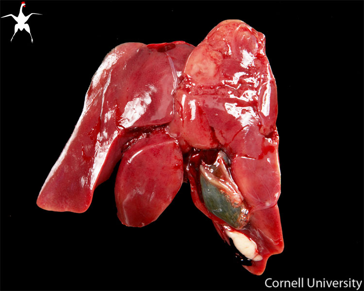
The gallbladder is located on the visceral surface of the right hepatic lobe.
It is normally dark green in color, due to the bile located within the lumen of this thin-walled structure. During autolysis, bile pigments may leak out of the gallbladder, staining the adjacent hepatic tissues yellow to green. This bile inbibition is a normal part of autolysis and should not be confused with a lesion. Similar staining can also occur in the ascending duodenum, adjacent to the area where the bile and pancreatic ducts empty. The size of the gallbladder is variable and may be enlarged in birds that are off-feed.
Record number:
10267
Case number:
Unknown
Age:
6 weeks
Breed:
White Leghorn
Clinical form:
Unknown
Infection type:
Unknown
Housing/mgmnt type:
Select One
Priority:
1
Rights:
© Cornell University
Etiology:
Tissues and organs:
Asset type:
Species:
Image:

