Submitted by admin on Sat, 09/20/2008 - 21:10
tissue_organ_import:
liver, spleen
Morphologic diagnosis:
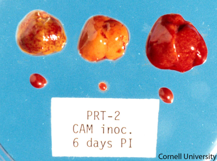
Liver and spleen (right): Hepato and splenomegaly
Spleen (left): Splenomegaly
Clinical description:
Enlarged, mottled, and congested liver and spleen collected from chicken embryos inoculated with *C. psittaci*. A normal liver and spleen are shown in the middle.
Pathologic description:
The livers and spleens from three embryos are depicted. The liver and the spleen in the center of the photo are normal. The other two sets of tissue are from embryos experimentally inoculated with *C. psittaci*. The liver and spleen on the right are enlarged. The spleen on the left is enlarged. Note: The mottled colors of the embryonic liver are often normal due to accumulation of lipid in the hepatocytes and areas of extramedullary hematopoiesis. Distinguishing these normal developmental changes from pathologic processes requires histopathology.
Record number:
7436
Case number:
Unknown
Clinical form:
Unknown
Infection type:
Experimental
Housing/mgmnt type:
Select One
Priority:
1
Image source URL:
http://cidc.library.cornell.edu/vet_avian/images/Chlam-Adjusted/CHLAM-053A.jpg
Etiology:
Exam findings:
Asset type:
Species:
Image:

