Submitted by admin on Sat, 09/20/2008 - 21:10
tissue_organ_import:
ileum, ceca
Clinical description:
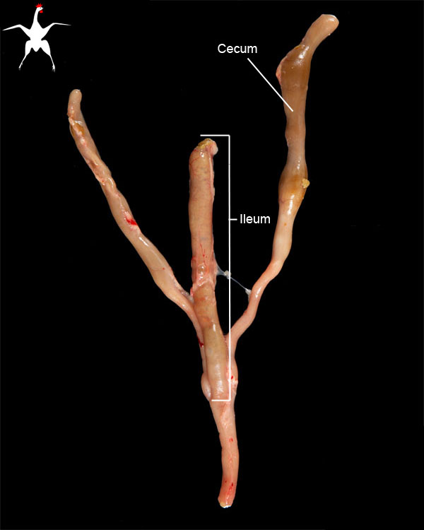
The serosa of the ileum should be shiny, tan, and smooth, with no thickening or bulges. Be careful not to over-interpret the color of the intestinal walls as post-mortem congestion and autolysis can quickly turn the intestinal walls red or black.
Because the intestinal walls are semi-translucent, look for areas of proliferation or mucosal exudate which can sometimes be visualized through the intestinal wall.
At the junction between the ileum and the descending colon, are two blind-ended sacs known as the ceca. In domestic poultry, the cecae are large structures that bend over themselves, with their apices pointing caudally. The walls should be thin and semi-translucent, allowing the greenish-colored intestinal contents to be visualized within. If the walls are opaque, thin or irregular, infection should be suspected.
Record number:
10242
Case number:
Unknown
Age:
6 weeks
Breed:
White Leghorn
Clinical form:
Unknown
Infection type:
Unknown
Housing/mgmnt type:
Select One
Priority:
1
Rights:
© Cornell University
Etiology:
Asset type:
Species:
Image:

