
Submitted by admin on Tue, 08/26/2008 - 15:40
tissue_organ_import:
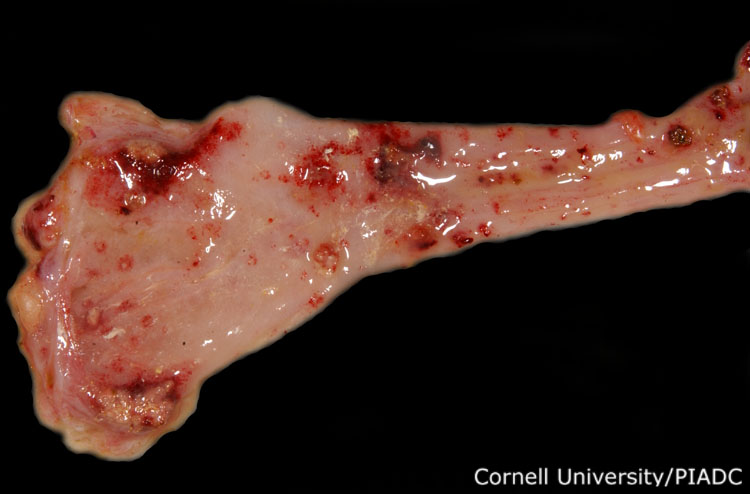
Colon, cloaca: ulcers, fibrin.
Morphologic diagnosis:
Colon and cloaca: Severe multifocal acute ulcers with fibrinous colitis and cloacitis
Clinical description:
This image was taken 5 days post experimental inoculation with viscerotropic velogenic Newcastle disease. Birds that survive the first several days of the infection typically have more severe post-mortem hemorrhagic lesions, as seen here.
Pathologic description:
The cloaca and the descending colon have been opened to reveal the mucosal surfaces. The mucosal surfaces are covered by multiple, randomly distributed, well demarcated defects that are covered by hemorrhage and slightly granular, pale yellow tags of deposits of tissue.
Record number:
21000
Case number:
5009
Age:
70 weeks
Breed:
White Leghorn SPF
Clinical form:
Unknown
Infection type:
Unknown
History:
The photograph of this bird was taken 4 days post inoculation. The bird was experimentally inoculated with Viscerotropic Velogenic Newcastle Disease virus [MO/31387/96] on 3/2/08 at Plum Island Animal Disease Center. The inoculation was performed via cloacal swab using 0.1ml.
Housing/mgmnt type:
Select One
Priority:
1
Rights:
© Cornell University
Etiology:
Exam findings:
Tissues and organs:
Asset type:
Species:
Image:
- Log in to post comments