
Submitted by admin on Tue, 08/26/2008 - 15:40
tissue_organ_import:
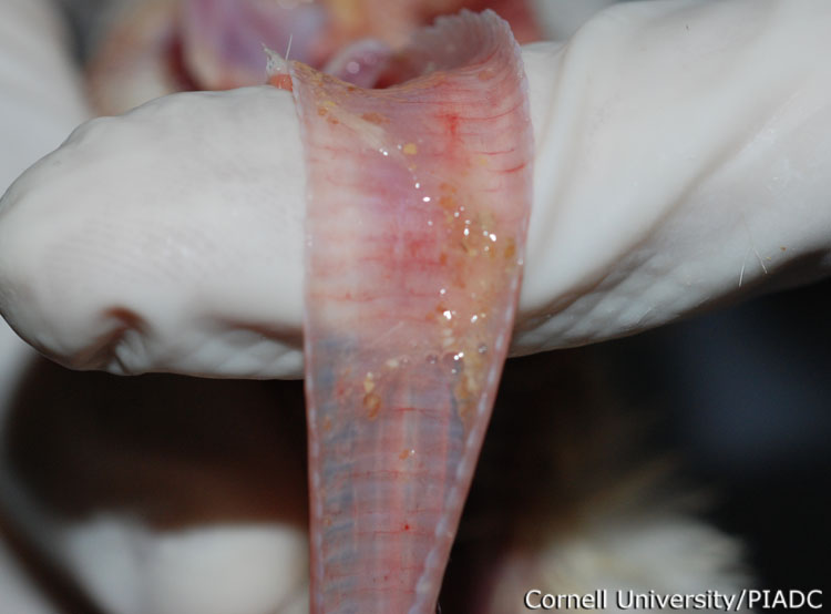
Trachea: petechia, catarrhal lesions
Morphologic diagnosis:
Trachea: Mild cattarhal tracheitis with petechia
Clinical description:
This image was taken 4 days post experimental inoculation with highly pathogenic avian influenza virus. There is mucopurulent catarrhal exudate in the lumen of the trachea.
Pathologic description:
The mucosal surface of the trachea is stippled by numerous pinpoint red foci and the lumen contains increased amounts of mucus.
Record number:
20948
Case number:
5030
Age:
16 weeks
Breed:
White Leghorn SPF
Clinical form:
Acute
Infection type:
Experimental
History:
The photograph was taken 4 days post inoculation. The bird was experimentally inoculated with highly pathogenic avian influenza virus on 3/2/08 at Plum Island Animal Disease Center. The inoculation was performed in the caudal thoracic air sac with strain A/CK/PA/469/3-84/H5N2, using 0.25ml.
Housing/mgmnt type:
Select One
Priority:
1
Rights:
© Cornell University
Etiology:
Exam findings:
Tissues and organs:
Asset type:
Species:
Image:
- Log in to post comments