
Submitted by admin on Tue, 08/26/2008 - 15:40
tissue_organ_import:
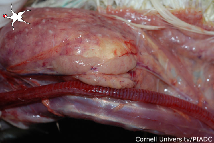
Trachea: hemorrhage, congestion; Thymus: atrophy.
Morphologic diagnosis:
Trachea: Acute diffuse mucosal hemorrhage. Thymus: Moderate atrophy.
Clinical description:
This image was taken 3 days post experimental inoculation with highly pathogenic avian influenza. There is severe diffuse hemorrhagic tracheitis which can be seen from the outer surface of the trachea. The thymus, adjacent to the trachea, also shows moderate atrophy in this 16 week old chicken.
Pathologic description:
This image shows a dissection of the neck. The trachea runs horizontally along the bottom of the picture. Immediately above the trachea is a thymic lobule. The trachea is closed, however the extensive reddening of the mucosa can be seen through the wall. Additionally, given the age of the chicken (16 weeks), the amount of thymic tissue is decreased.
Record number:
20916
Case number:
5048
Age:
16 weeks
Breed:
White Leghorn SPF
Clinical form:
Acute
Infection type:
Experimental
History:
The photograph was taken 3 days post inoculation. The bird was experimentally inoculated with highly pathogenic avian influenza virus on 3/2/08 at Plum Island Animal Disease Center. The inoculation was performed in the caudal thoracic air sac with strain A/CK/PA/469/3-84/H5N2, using 0.25ml.
Housing/mgmnt type:
Select One
Priority:
1
Etiology:
Tissues and organs:
Asset type:
Species:
Image:
- Log in to post comments