
Submitted by admin on Tue, 08/26/2008 - 15:40
tissue_organ_import:
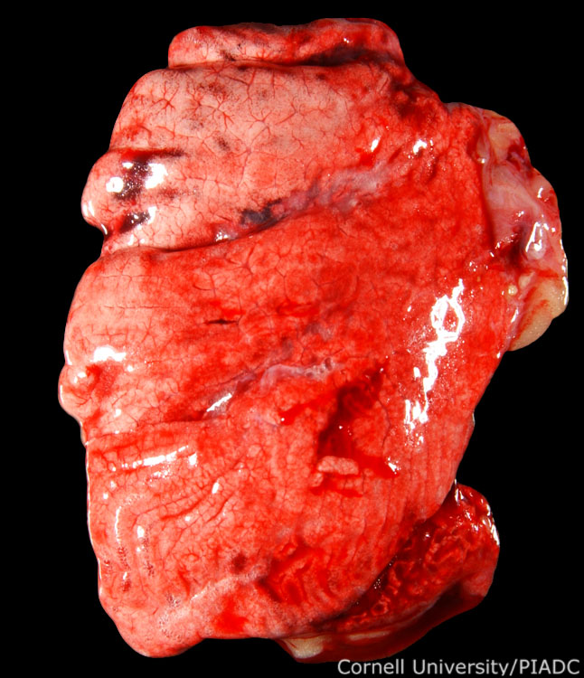
Lungs: congestion, edema, hemorrhage.
Morphologic diagnosis:
Lung: Acute multifocal hemorrhage with edema and congestion
Clinical description:
This image was taken 2 days post experimental inoculation with highly pathogenic avian influenza. As commonly observed in HPAI, the lungs are swollen, due to edema, congested, and there are extensive hemorrhages throughout the lungs.
Pathologic description:
There are several, irregularly shaped red foci in the lungs. The entire pulmonary parenchyma is wet and glistening with some areas of reddening. The blood vessels and intralobular spaces are prominent.
Record number:
20796
Case number:
5005
Age:
70 weeks
Breed:
White Leghorn SPF
Clinical form:
Acute
Infection type:
Experimental
History:
The photograph was taken 2 days post inoculation. The bird was experimentally inoculated with highly pathogenic avian influenza virus on 3/2/08 at Plum Island Animal Disease Center. The inoculation was performed in the caudal thoracic air sac with strain A/CK/PA/469/3-84/H5N2, using 0.25ml.
Housing/mgmnt type:
Select One
Priority:
1
Rights:
© Cornell University
Etiology:
Exam findings:
Tissues and organs:
Asset type:
Species:
Image:
- Log in to post comments