
Submitted by admin on Tue, 08/26/2008 - 15:40
tissue_organ_import:
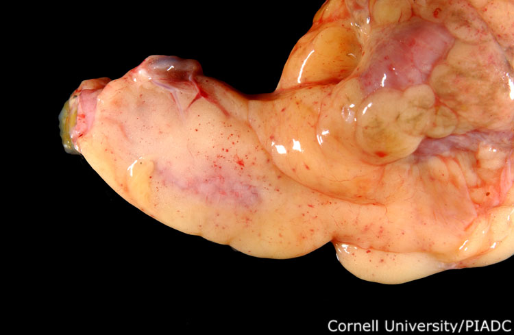
Coelomic cavity, serosal fat: petechiae.
Morphologic diagnosis:
Coelomic fat: Mild acute multifocal petechiation
Clinical description:
This image was taken 3 days post experimental inoculation with highly pathogenic avian influenza. In HPAI, many small petechiae will often cover the abdominal fat and serosal surfaces of many organs, as with the surface of this proventriculus.
Pathologic description:
The adipose tissue surrounding the coelomic organs is covered by numerous distinct pinpoint red foci.
Record number:
20868
Case number:
5004
Age:
70 weeks
Breed:
White Leghorn SPF
Clinical form:
Acute
Infection type:
Experimental
History:
The photograph was taken 3 days post inoculation. The bird was experimentally inoculated with highly pathogenic avian influenza virus on 3/2/08 at Plum Island Animal Disease Center. The inoculation was performed in the caudal thoracic air sac with strain A/CK/PA/469/3-84/H5N2, using 0.25ml.
Housing/mgmnt type:
Select One
Priority:
1
Rights:
© Cornell University
Etiology:
Exam findings:
Tissues and organs:
Asset type:
Species:
Image:
- Log in to post comments