
Submitted by admin on Tue, 08/26/2008 - 15:40
Clinical Signs:
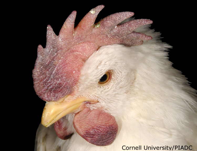
Edema (comb, wattles, eyelids, periorbital); Ecchymoses (comb, wattles)
Morphologic diagnosis:
Subcutaneous tissue (comb, wattles): Severe acute multifocal hemorrhage with edema. Subcutaneous tissue (face, eyes): Moderate edema
Clinical description:
This image was taken 3 days post experimental inoculation with highly pathogenic avian influenza. The degree of edema, hemorrhages, and necrosis present on the comb and wattles will depend on the stage of this disease. Here, severe edema and hemorrhages are observed.
Pathologic description:
Within the subcutaneous tissues of the comb and wattles there are large, poorly demarcated dark red areas. The wattles are slightly swollen and the periocular and eyelid skin is swollen.
Record number:
20812
Case number:
5045
Age:
16 weeks
Breed:
White Leghorn SPF
Clinical form:
Acute
Infection type:
Experimental
History:
The photograph was taken 3 days post inoculation. The bird was experimentally inoculated with highly pathogenic avian influenza virus on 3/2/08 at Plum Island Animal Disease Center. The inoculation was performed in the caudal thoracic air sac with strain A/CK/PA/469/3-84/H5N2, using 0.25ml.
Housing/mgmnt type:
Select One
Priority:
1
Rights:
© Cornell University
Etiology:
Exam findings:
Tissues and organs:
Asset type:
Species:
Image:
- Log in to post comments