
Submitted by admin on Tue, 08/26/2008 - 15:40
tissue_organ_import:
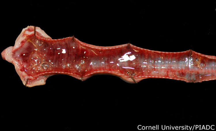
Trachea: hemorrhages, catarrhal lesions.
Morphologic diagnosis:
Trachea: Severe acute cattarhal tracheitis with hemorrhage
Clinical description:
This image was taken 3 days post experimental inoculation with highly pathogenic avian influenza. In HPAI, the upper respiratory tract may have catarrhal lesions including fibrinous, serofibrinous, mucopurulent (seen here), or fibrinopurulent exudates.
Pathologic description:
The mucosa of the trachea is dark red and there are numerous coalescing dark red areas throughout the wall. The tracheal lumen is filled with increased amounts of mucus.
Record number:
20900
Case number:
5027
Age:
16 weeks
Breed:
White Leghorn SPF
Clinical form:
Acute
Infection type:
Experimental
History:
The photograph was taken 3 days post inoculation. The bird was experimentally inoculated with highly pathogenic avian influenza virus on 3/2/08 at Plum Island Animal Disease Center. The inoculation was performed in the caudal thoracic air sac with strain A/CK/PA/469/3-84/H5N2, using 0.25ml.
Housing/mgmnt type:
Select One
Priority:
1
Rights:
© Cornell University
Etiology:
Exam findings:
Tissues and organs:
Asset type:
Species:
Image:
- Log in to post comments