
Submitted by admin on Tue, 08/26/2008 - 15:40
tissue_organ_import:
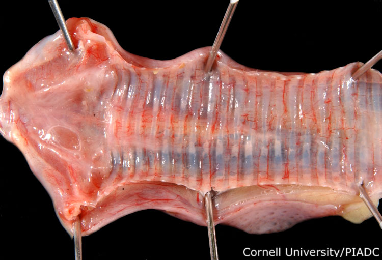
Trachea: congestion.
Morphologic diagnosis:
Tracheal mucosa: Mild congestion
Clinical description:
This image was taken 2 days post experimental inoculation with highly pathogenic avian influenza. The range of tracheal lesions observed may vary greatly from mucous accumulation to severe hemorrhagic tracheitis. Here, mild congestion of the tracheal mucosa can be observed.
Pathologic description:
The trachea has been opened to reveal the mucosal surface. The blood vessels are prominent and congested.
Record number:
20754
Case number:
5005
Age:
70 weeks
Breed:
White Leghorn SPF
Clinical form:
Acute
Infection type:
Experimental
History:
The photograph was taken 2 days post inoculation. The bird was experimentally inoculated with highly pathogenic avian influenza virus on 3/2/08 at Plum Island Animal Disease Center. The inoculation was performed in the caudal thoracic air sac with strain A/CK/PA/469/3-84/H5N2, using 0.25ml.
Zootechnical purpose:
Egg layers- white egg
Housing/mgmnt type:
Select One
Priority:
1
Rights:
© Cornell University
Etiology:
Exam findings:
Tissues and organs:
Asset type:
Species:
Image:
- Log in to post comments