
Submitted by admin on Tue, 08/26/2008 - 15:40
tissue_organ_import:
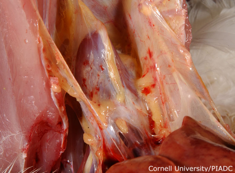
Pericardium and pericardial fat: petechia.
Morphologic diagnosis:
Pericardium and adipose tissue: Multifocal acute petechiae.
Clinical description:
This image was taken 2 days post experimental inoculation with highly pathogenic avian influenza. With most strains for HPAI, as seen here, pinpoint petechial hemorrhages are frequently observed along the pericardium and abdominal fat.
Pathologic description:
The keel and rib cage have been cut and reflected cranially. The heart has remained attached to the ventral surface of the sternum and has been pulled forward as well. The apex of the heart is at the top of the image and the liver is at the bottom right. The pericardial surface and the surrounding adipose tissue is stippled with small, discrete red foci.
Record number:
20776
Case number:
5005
Age:
70 weeks
Breed:
White Leghorn SPF
Clinical form:
Acute
Infection type:
Experimental
History:
The photograph was taken 2 days post inoculation. The bird was experimentally inoculated with highly pathogenic avian influenza virus on 3/2/08 at Plum Island Animal Disease Center. The inoculation was performed in the caudal thoracic air sac with strain A/CK/PA/469/3-84/H5N2, using 0.25ml.
Housing/mgmnt type:
Select One
Priority:
1
Rights:
© Cornell University
Etiology:
Exam findings:
Tissues and organs:
Asset type:
Species:
Image:
- Log in to post comments