
Submitted by admin on Tue, 08/26/2008 - 15:40
tissue_organ_import:
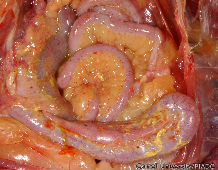
Intestines and coelomic cavity: petechiae.
Morphologic diagnosis:
Coelomic cavity: Moderate acute multifocal petechia. Coelomic cavity: Fibrinous coelomitis (presumed secondary to unrelated egg-yolk peritonitis)
Clinical description:
This image was taken 2 days post experimental inoculation with highly pathogenic avian influenza. In HPAI, pinpoint petechial lesions are common on the serosal surfaces of organs such as the intestines, seen here.
Pathologic description:
The serosal surfaces of the intestinal loops and the mesenteric fat are stippled by numerous, pinpoint red foci and portions of the intestines are coated by strands of yellow, friable material.
Record number:
20782
Case number:
5005
Age:
70 weeks
Breed:
White Leghorn SPF
Clinical form:
Acute
Infection type:
Experimental
History:
The photograph was taken 2 days post inoculation. The bird was experimentally inoculated with highly pathogenic avian influenza virus on 3/2/08 at Plum Island Animal Disease Center. The inoculation was performed in the caudal thoracic air sac with strain A/CK/PA/469/3-84/H5N2, using 0.25ml.
Housing/mgmnt type:
Select One
Priority:
1
Rights:
© Cornell University
Etiology:
Exam findings:
Tissues and organs:
Asset type:
Species:
Image:
- Log in to post comments