
Submitted by admin on Tue, 08/26/2008 - 15:40
tissue_organ_import:
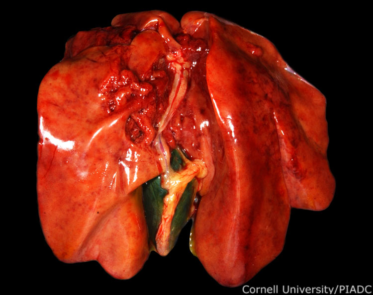
Liver: hemorrhages.
Morphologic diagnosis:
Liver: Multifocal acute necrosis with hemorrhage
Clinical description:
This image was taken 2 days post experimental inoculation with highly pathogenic avian influenza. In HPAI, it is common to find hemorrhagic lesions distributed throughout visceral organs. Here, hemorrhages on found on the surface of the liver. Occasionally, the liver may also have necrotic foci.
Pathologic description:
The liver is mottled by multiple, often coalescing red foci. On the right edge of the image, near the junction of the two portions of the left liver lobe, these red foci appear slightly sunken. The remaining parenchyma is slightly pale and during the examination, the tissue was observed to be friable.
Record number:
20781
Case number:
5005
Age:
70 weeks
Breed:
White Leghorn SPF
Clinical form:
Acute
Infection type:
Experimental
History:
The photograph was taken 2 days post inoculation. The bird was experimentally inoculated with highly pathogenic avian influenza virus on 3/2/08 at Plum Island Animal Disease Center. The inoculation was performed in the caudal thoracic air sac with strain A/CK/PA/469/3-84/H5N2, using 0.25ml.
Housing/mgmnt type:
Select One
Priority:
1
Rights:
© Cornell University
Etiology:
Exam findings:
Tissues and organs:
Asset type:
Species:
Image:
- Log in to post comments